Is something wrong right now? If your pet is struggling to breathe, has collapsed, is bleeding heavily, cannot pass urine, has a swollen hard abdomen, is having a seizure, has been hit by a car, or has swallowed something you think is poisonous, this is an emergency. Call us before you set off so we can be ready for you, and if we are closed call a 24-hour emergency hospital straight away. Do not wait to see whether it settles. Call 000-000-0000
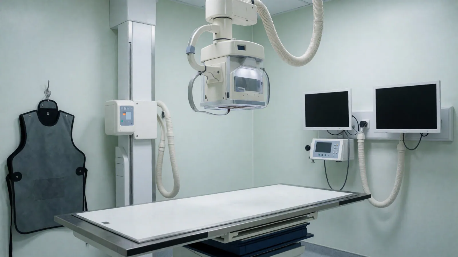
A radiograph and an ultrasound scan are not two versions of the same picture — they answer genuinely different questions, and part of our job is working out which one your animal actually needs before either machine is switched on. A digital x-ray shows shape, position and density: whether a bone is broken, where a swallowed object has stopped, whether a chest looks the way a chest should. An ultrasound shows texture and structure inside a soft organ that a radiograph renders as a single grey shadow — whether a kidney's internal architecture looks normal, whether there is fluid where there should not be any at all.
Sedation is not used by default, and most conscious animals lie still enough for a routine limb or chest radiograph without it. It becomes the right choice when an animal is too anxious, in too much pain, or simply will not hold the specific position a diagnostic image needs, and forcing a frightened animal to stay still for repeated attempts produces poor images and a worse experience than a short, monitored sedation would. Every sedation is assessed individually against the animal in front of us, dosed accordingly, and monitored throughout — never applied as a blanket policy to save time or effort.
You will not be in the room for either study, and the reason is straightforward rather than bureaucratic. Radiography uses ionising radiation, and repeated low-dose exposure to a person holding an animal in position, appointment after appointment, is a real occupational risk we are not willing to create for you or for our own staff. Ultrasound has no radiation risk at all, but an animal that is anxious about your absence in the room is frequently calmer without you there than people expect, and a calmer animal produces a scan that takes less time overall to complete.
What a radiograph answers that an ultrasound cannot, and the other way round
Each modality is built to answer a specific kind of question, and reading the wrong one for the job wastes time and your money.
A broken or displaced bone
A digital radiograph is still the fastest, clearest way to confirm a fracture, show exactly how the bone ends line up, and check a healing bone against an earlier image at a recheck. Ultrasound cannot see through bone at all, so a suspected fracture is always a radiograph question, never an ultrasound one, regardless of which machine happens to be free first.
Where a swallowed object has stopped
Many swallowed objects — coins, small toys, some fabric — show clearly on a radiograph because of their density, and a series of views can often show whether an object is moving through the gut or stuck in one place. Some materials are radiolucent and do not show up this way, in which case an ultrasound or a different study is proposed instead, and we will tell you plainly which situation applies.
Whether an organ is the wrong size or position
A radiograph shows the outline, size and position of the abdominal organs as shadows against each other, which is enough to flag an obviously enlarged liver, a displaced bladder, or a mass pushing other organs out of their normal place. It is a silhouette, though — it cannot tell you what that organ's own tissue looks like on the inside, which is precisely where ultrasound takes over from there.
What an organ's own tissue actually looks like
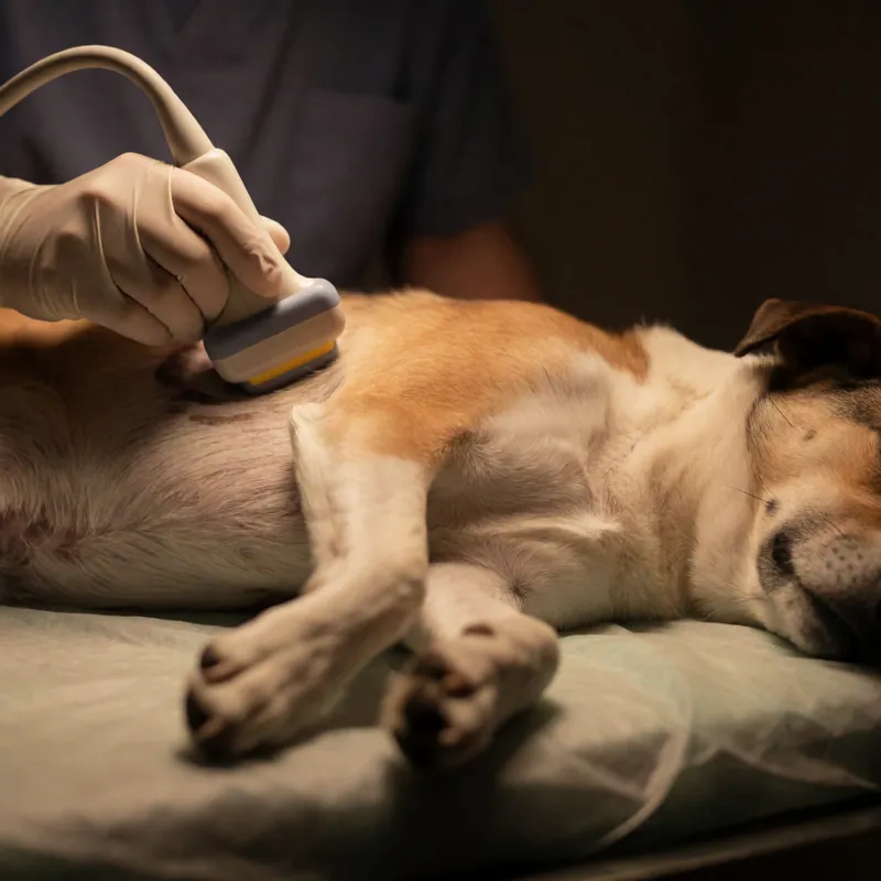
Ultrasound builds a live image from sound waves reflecting off tissue, which shows texture and internal structure that a radiograph simply cannot resolve — whether a kidney has lost its normal internal pattern, whether a mass is solid or fluid-filled, whether an organ's wall is thickened. This is the question a radiograph physically cannot answer, no matter how many views are taken of it.
Fluid where there should not be any
Free fluid in the abdomen or around the heart is often invisible or ambiguous on a radiograph, appearing as a general loss of detail rather than a clear finding, whereas ultrasound shows it immediately and lets us estimate roughly how much is there and often guide a needle to sample it safely. This single difference is frequently the reason we move from one machine to the other within the same visit.
Sedation — why, and why not always
Sedation is a clinical decision made about the animal in front of us, not a routine step in every imaging appointment.
Most routine radiographs need none at all
A calm dog or cat can usually be positioned for a standard limb or chest x-ray by a technician using gentle handling and a few seconds of stillness, and the large majority of routine radiographs here are taken this way. Sedation is added when it is actually needed, not offered as a default for convenience or speed on a busy day.
When a position genuinely cannot be held awake
Certain radiographic views require an animal to lie in a position that is uncomfortable or unnatural for a few seconds longer than a conscious, anxious animal will tolerate — a hip view for suspected dysplasia is the clearest example. Repeated failed attempts produce blurred, non-diagnostic images and a distressing appointment, so a short sedation is the kinder and more accurate choice here.
Pain changes the calculation entirely
An animal in genuine pain, such as a suspected fracture, cannot and should not be asked to hold a position through discomfort, and attempting it without sedation is neither humane nor likely to produce a usable image. Sedation combined with pain relief is standard in these cases and is assessed and dosed for the individual animal in front of us that day.
Ultrasound almost never needs sedation
Ultrasound requires an animal to lie reasonably still on a padded table in a dimmed room for several minutes, which most cats and dogs manage without sedation once they have settled, particularly with a technician supporting them gently throughout. Sedation for ultrasound is the exception, usually reserved for an animal that is especially anxious or in pain from an unrelated cause.
Monitoring during any sedation
Any sedated animal is monitored for heart rate, breathing and colour throughout the procedure and during recovery afterward, by a technician whose attention is on that animal specifically for that hour. You will get a full account of what was given and why, and sedation is never added to an estimate as an unexplained line item on the bill. Most animals are steady enough to go home within an hour or two, and we will not release your animal to you until we are satisfied it is genuinely ready.
The appointment itself, and why you wait outside
Both studies happen in a dimmed, quiet room, and neither one needs you standing beside the table.
Why radiation safety keeps owners out of the room
Digital radiography uses a genuinely low dose of ionising radiation, safe for an occasional patient but not for a person holding animals in position appointment after appointment, day after day. That accumulated exposure is a real occupational risk, which is why our own technicians use positioning aids and protective equipment rather than hands, and why you are asked to wait just outside rather than assist.
Ultrasound has no radiation risk, but the room still works better without you
There is no dose consideration for ultrasound, but an anxious animal is often noticeably calmer without its owner present than people expect, because some of what reads as separation distress is actually the animal reading tension in you. A technician who handles anxious animals daily can usually settle one faster alone than with a worried owner in the room. The exception is a specific request from your own veterinarian, which we will always honour if it is clinically reasonable to do so.
Images kept against your animal's record
Every radiograph and ultrasound image is stored digitally against your animal's file, not handed to you on a disc and forgotten, so a healing fracture, a growing mass or a kidney's appearance over several years can be compared image to image rather than described from memory at each visit that follows. This matters most for anything being watched over time rather than treated once and closed, where the comparison between this year's image and last year's is often more informative than either image looked at on its own.
Results explained the same visit
A veterinarian reviews the images with you before you leave wherever that is possible, in plain language, showing you on screen what was found rather than reading a report aloud. Where a second opinion or a specialist radiologist's reading is genuinely useful for a complex case, we will say so and arrange it for you. You are welcome to ask us to point to the specific area on the image we are describing, and to take a phone photo of the screen for your own records if that helps you remember the conversation later.
What happens if something urgent turns up mid-scan
Occasionally an ultrasound or radiograph finds something that changes the whole visit — a mass, free fluid, an obstruction — and when that happens we stop, call you if you are not already in the building, and talk through the finding and the options before anything further is done. An imaging finding is information, not an automatic decision to act.
X-Ray & Ultrasound in western Queens
Digital radiography for a bone, an object or an organ's size and position, and ultrasound for what that organ's own tissue looks like on the inside, with images kept on your animal's record.
It starts the same way whatever you are here for: a full examination with time to talk, what we found explained in plain English, and a written estimate for anything beyond the consultation before it happens. You are entitled to take that estimate away and think about it, and to ask what happens if you do nothing. That last one is a real question and it gets a real answer.
This is general information, not veterinary advice. This page describes a service. It is general information and it is not veterinary advice, a diagnosis or a treatment plan for any particular animal, because no page can examine your pet. If your animal is unwell, injured or behaving unusually, speak to a licensed veterinarian who can actually see it. In an emergency contact your own veterinarian or a 24-hour emergency animal hospital immediately.
Questions
X-Ray & Ultrasound — the questions we are asked most
Does my pet need an x-ray or an ultrasound?
It depends entirely on the question we are trying to answer. A radiograph is the right tool for a suspected fracture, a swallowed object, or checking the size and position of the abdominal organs. An ultrasound is the right tool for looking at what an organ's own tissue looks like on the inside, or finding fluid that a radiograph cannot show clearly. We will examine your animal first and tell you which study actually answers the question.
Will my pet need to be sedated for an x-ray?
Often not. Most calm animals can be positioned for a routine limb or chest radiograph with gentle handling alone, and sedation is added only when a specific position cannot be held comfortably while awake, when an animal is too anxious, or when pain makes holding still unreasonable to ask. Every sedation is assessed and dosed for the individual animal and monitored throughout, and we will explain why it is being recommended before you agree.
Why can't I stay in the room during the x-ray?
Digital radiography uses a genuinely low but real dose of ionising radiation, and while an occasional exposure for a patient is not a concern, repeated exposure for a person holding animals in position appointment after appointment is a real occupational risk we will not create for you. Our technicians use positioning aids and protective equipment instead of hands wherever possible. Ultrasound carries no radiation risk, but most anxious animals are calmer without an owner present.
How much does a pet x-ray cost in Woodside, NY?
The first view is $180 and includes positioning and a sedation assessment. Additional views taken on the same visit, which are common for a full survey of a limb or a chest, are $65 each. An abdominal ultrasound is a separate study at $320. We will tell you before the appointment roughly how many views your animal's situation is likely to need, though the final number sometimes depends on what the first image shows.
What does an abdominal ultrasound show that an x-ray does not?
An ultrasound shows the internal texture and structure of an organ — whether a kidney's internal pattern looks normal, whether a mass is solid or filled with fluid, whether an organ wall is thickened — none of which a radiograph can resolve, because a radiograph only shows an organ's outline and position as a shadow. The two studies are frequently used together, a radiograph first, then an ultrasound to look closely at one area of concern.
Is sedation safe for an older or sick animal?
Sedation is assessed against the specific animal in front of us, not applied as a blanket policy, and an older or unwell animal often needs a different drug choice and a lower dose than a young healthy one — which is exactly why the assessment happens every time rather than being skipped for a routine case. Heart rate, breathing and colour are monitored throughout and during recovery, and we will talk you through the plan first.
How long does it take to get the results?
Images are reviewed the same visit in almost every case, and a veterinarian will talk you through what was found in plain language before you leave, showing you the image on screen rather than simply reading out a report. Where a case is genuinely complex enough to benefit from a specialist radiologist's second reading, we will say so and arrange it, and let you know honestly that the full answer will take a little longer.
Will you keep my pet's old x-rays and ultrasound images?
Yes, every image is stored digitally against your animal's own record rather than handed to you once and forgotten, which means a healing fracture, a slow-growing mass or a kidney's appearance over several years can be compared directly, image to image, at a later visit. If you are transferring to another practice, we will send the images along with the rest of the records, free, the same working day.
Related
Usually booked alongside this
Diagnostics & Medicine
Sick Visits
Same-day appointments held back on every day of the week for an animal that was well yesterday and is not well today, with a desk trained to tell how quickly you need to be seen.
Read more
Diagnostics & Medicine
In-House Laboratory
Haematology, biochemistry, urinalysis and cytology run in the building, with most results back inside the hour — the difference between starting treatment today and starting it after a weekend wait.
Read more
Diagnostics & Medicine
Skin, Ear & Allergy
Itch is a symptom rather than a diagnosis, and the same red, irritated ear or paw can have several different causes that a cytology sample taken at the visit narrows down before anything is prescribed.
Read moreA written estimate before anything is booked
Book an appointment
We hold same-day appointments back on every single day of the week for animals that were well yesterday. Call the practice and describe what you have noticed, and the desk will book you at the right speed rather than the soonest one.